
Radiography is the main imaging modality for the assessment of bone and bone structure. TRH Equine Veterinary Services uses the very latest in digital computer radiography systems which produces radiographic images of excellent quality. The pictures are viewed on a computer screen and areas of particular interest can be focussed on to achieve more detail. This system allows us to easily send digital copies of your xrays. The system we have is fully portable allowing us to obtain high quality xray images at your property.
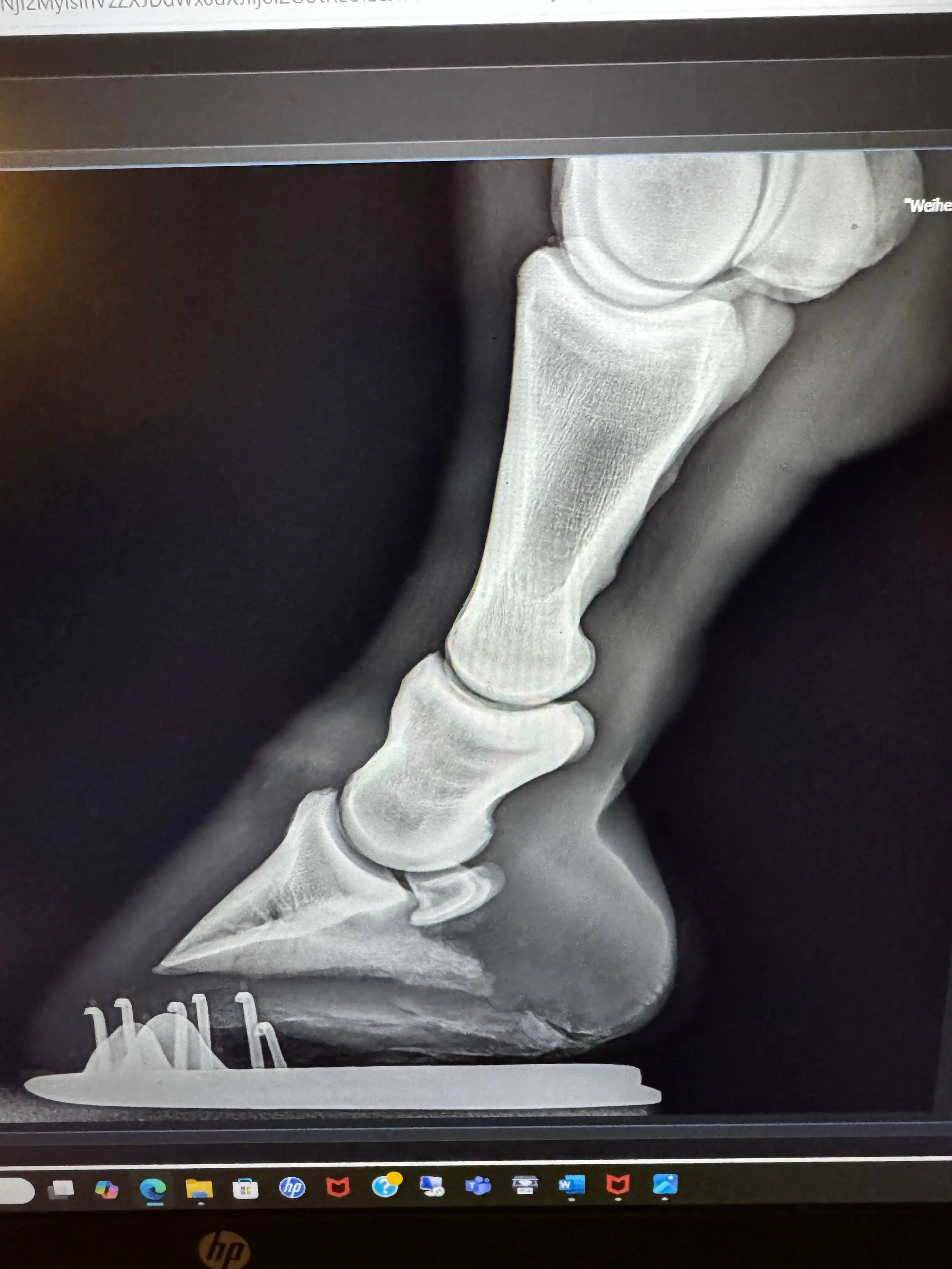
We have a number of high quality portable X-ray machines. As long as we have power and a safe, dry area we can perform radiograph anywhere. Good foot radiographs require a flat hard surface. Foot radiographs allow us to work with the farrier to prevent and treat lameness by assessing each horse and each foot as an individual. Hind feet can be just as important as front feet angles and can even help address back and hock pain.
Frequently asked questions
Ultrasonography is an imaging modality used to assess soft tissue structures such as tendons, ligaments, internal organs, eyes and masses. Damage to tendons and ligaments can be seen on ultrasound as changes to the fibre pattern and cross sectional area of the structure.
Ultrasound is used rectally in mares to assess the uterus and ovaries as well as checking for pregnancy during their breeding cycle. TRH Equine Veterinary Services has a high quality soft tissue ultrasound as well reproduction ultrasound for mobile use, allowing us to perform thorough ultrasound exams in the stables and field.
Mares who behave erratically or exhibit stallion like behaviour or seem constantly in season may have a reproductive condition such as anovulatory follicles or granulosa thecal cell tumours. These can be diagnosed by rectal ultrasound though a crush or similar restraint is needed to prevent injury to the mare or vet! Hormones can also be assessed via blood tests. Ultrasound can also be used to investigate lumps and swelling anywhere on the body. It is very useful over the pelvis and upper limbs to check for bone injuries where X-ray cannot penetrate. Ultrasound guided injections are often used for stifles and sacroiliac joints.